
That dark, leathery crust covering a non-healing wound isn't a protective scab; it's a clinical mask that hides the true extent of tissue damage. While a scab is a sign of healthy recovery, necrotic eschar is dead tissue that acts as a physical barrier, preventing the body from repairing itself and often trapping dangerous bacteria underneath. It's completely normal to feel a sense of dread when you notice a wound turning black or hard, especially if you're caring for an elderly family member who can't easily travel to a specialist. The fear of deep-seated infection or the looming threat of amputation is a heavy burden for any caregiver to carry alone.
In this guide, you'll learn how to accurately identify necrotic tissue, recognize the subtle signs of infection, and understand why professional intervention is vital for recovery. We'll also discuss how advanced mobile wound care provides a safer, more convenient alternative to traditional clinic visits. We'll explore the clinical risks of leaving dead tissue untreated and explain how bedside evaluations from board-certified specialists bring expert regenerative medicine directly to your home, ensuring that your loved one receives hospital-grade care without the stress of transport.
Key Takeaways
- Identify the clinical characteristics of necrotic eschar and learn why this leathery tissue is a barrier to healing rather than a protective layer.
- Understand the risks associated with unstageable wounds and how professional debridement reveals the true severity of an injury.
- Compare the differences between superficial scabs and deep tissue necrosis to better monitor wound progression at home.
- Discover how board-certified specialists provide bedside evaluations and advanced debridement services throughout Glendale and Los Angeles County.
- Recognize why self-treatment of necrotic tissue is hazardous and how clinical intervention prevents hospitalizations and serious complications.
What is Eschar? Defining Necrotic Tissue in Chronic Wounds
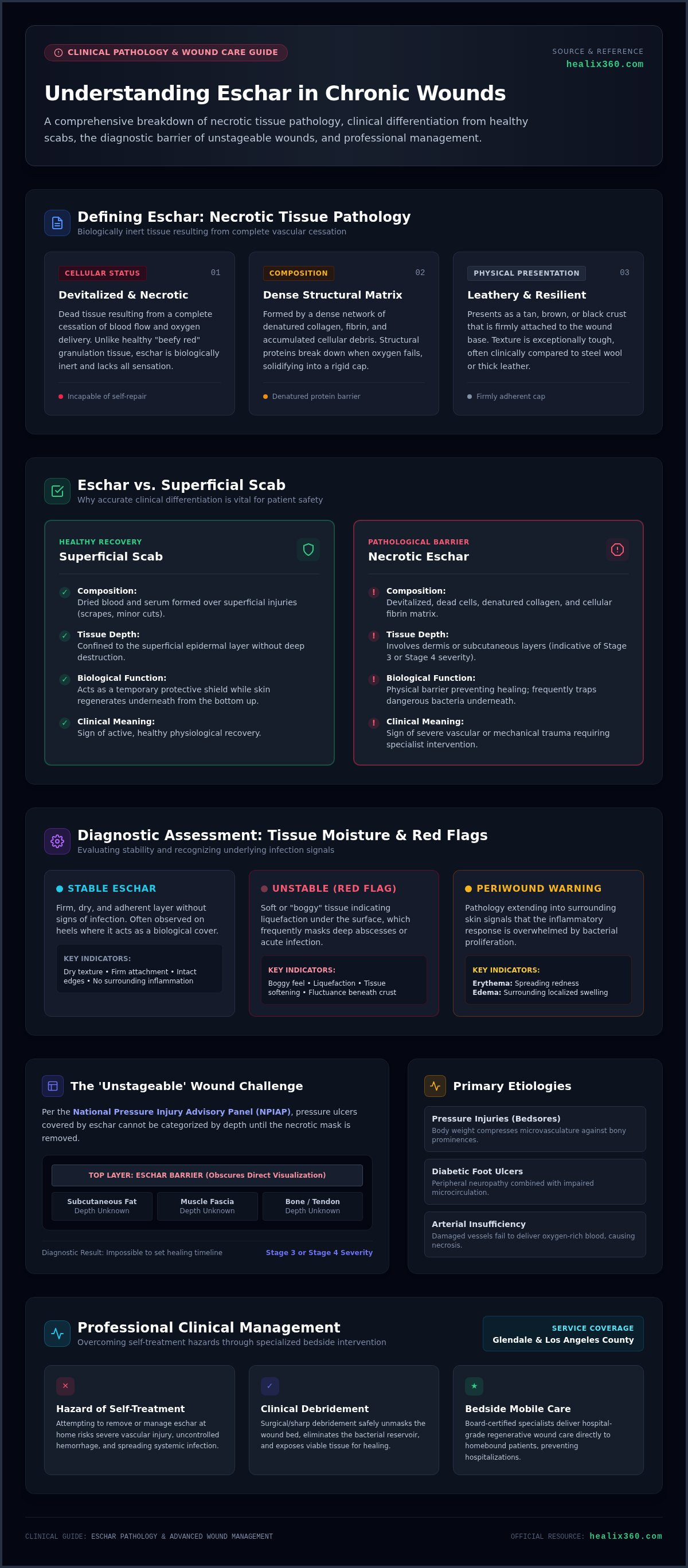
In a clinical setting, eschar is defined as a collection of devitalized, necrotic cells that have accumulated within a wound bed. This tissue is essentially dead. It's the direct result of a complete cessation of blood flow and oxygen to a specific area of the body. While healthy granulation tissue is characterized by a "beefy red" appearance and a moist environment conducive to healing, necrotic tissue is biologically inert. It typically presents as a tan, brown, or black leathery covering that lacks any sensation. This material is composed of a dense matrix of denatured collagen, fibrin, and cellular debris. When oxygen delivery fails, these structural proteins begin to break down, creating the rigid, non-viable cap we see at the surface. We frequently observe this in patients suffering from arterial ulcers or Stage 3 and 4 pressure injuries where prolonged ischemia has caused significant tissue death.
Identifying Eschar: Texture and Appearance
Specialists often identify this tissue by its distinct texture, which can feel as tough as steel wool or thick leather. It's remarkably resilient and often firmly attached to the wound base. The moisture level of this tissue tells us a great deal about the wound's status. Firm, dry tissue is often described as "stable," whereas soft or boggy areas suggest that the tissue is undergoing liquefaction. This softening is a major red flag, as it often masks a deep-seated infection or an abscess that requires immediate clinical attention. We also carefully evaluate the periwound area for erythema and edema. These signs of redness and swelling indicate that the inflammatory process is extending beyond the necrotic site, which may suggest that the body is struggling to manage a high bacterial load or a spreading infection.
Eschar vs. Scab: Why the Difference Matters
The distinction between a common scab and necrotic tissue is a critical one for patient safety. A scab consists of dried blood and serum that forms over a superficial injury, such as a scrape or a minor cut. It acts as a temporary protective barrier while the skin underneath regenerates from the bottom up. In contrast, eschar represents a deeper level of destruction that involves the dermis or even subcutaneous layers. It's not a sign of healthy recovery; it's a sign of significant vascular or mechanical trauma that has killed the underlying tissue. Mistaking necrotic tissue for a simple scab leads to a dangerous delay in treatment that allows underlying infections to proliferate and tissue damage to deepen.
Causes of Eschar and the 'Unstageable' Wound Challenge
The primary driver of necrotic tissue formation is prolonged ischemia. When blood flow to the skin and underlying structures is restricted for an extended period, cells die from oxygen deprivation. This process is most frequently observed in pressure injuries, commonly known as bedsores, where the weight of the body compresses small blood vessels against a bony prominence. Diabetic foot ulcers also present a high risk due to a combination of peripheral neuropathy and impaired microcirculation. In the lower extremities, arterial insufficiency is a major contributor, as damaged vessels cannot deliver enough nutrient-rich blood to maintain tissue viability. Because eschar involves the death of deep tissue layers, its presence is a definitive clinical indicator that the wound has progressed to Stage 3 or Stage 4 severity.
Why Your Wound is Classified as 'Unstageable'
According to the National Pressure Injury Advisory Panel (NPIAP), unstageable pressure ulcers covered by eschar cannot be accurately categorized until the necrotic material is removed. The dark, leathery layer acts as a physical mask that obscures the wound bed. A clinician cannot determine if the injury has reached the fat, muscle, or bone while this barrier remains in place. This lack of visibility creates a diagnostic challenge, making it impossible to develop a precise treatment plan or predict a healing timeline. Removing this mask through professional debridement is a clinical necessity to reveal the true depth of the injury and initiate the recovery process.
Stable vs. Unstable Eschar
While most necrotic tissue requires removal, there is a specific clinical exception known as "stable eschar." This typically occurs on the heels, where a dry, intact, and firm layer may act as the body's natural cover. If the tissue remains adherent and shows no signs of inflammation, specialists may choose to leave it undisturbed to protect the underlying structures. However, you must monitor for signs of "unstable" tissue. If the area becomes boggy, develops a foul odor, or begins to drain fluid, the tissue has become a reservoir for infection. These changes require an urgent professional assessment to prevent systemic complications. Our specialists provide bedside wound evaluations to help families determine if debridement is necessary without the risk of transporting a vulnerable patient.
Eschar vs. Scabs: A Comparison for Patients and Caregivers
Distinguishing between a common scab and necrotic tissue is essential for proper wound management. While they may look similar to the untrained eye, their biological origins and risks are vastly different. A scab is a temporary crust formed by dried blood and serum following a superficial injury to the epidermis. In contrast, eschar represents deep tissue death that often extends through the dermis and into subcutaneous layers. Proper wound assessment and necrotic tissue classification allow clinicians to differentiate these structures and determine the necessary level of intervention.
The following points highlight the primary differences between these two types of tissue:
- Biological Origin: Scabs form from external fluids drying on the surface; necrotic tissue forms from internal cellular death.
- Attachment: Scabs eventually develop loose, curling edges as healthy skin heals underneath. Necrotic tissue remains tightly integrated into the wound bed, often appearing flush with or even recessed into the surrounding skin.
- Healing Trajectory: Healthy skin pushes a scab off naturally once recovery is complete. Necrotic tissue rarely sheds on its own and typically requires professional debridement to allow for granulation.
- Risk Profile: Scabs are generally low-risk indicators of healing. Necrotic tissue carries a high risk of masking an underlying abscess or a deep-seated infection that can lead to systemic complications.
When to Worry: Warning Signs of Infected Eschar
Caregivers must remain vigilant for signs that necrotic tissue has become unstable or infected. One of the most concerning indicators is a "boggy" or fluctuant texture. This suggests that the hard surface is actually floating on a pocket of pus or fluid. You should also watch for systemic symptoms such as fever, chills, or a sudden increase in localized pain. If the skin surrounding the site becomes hot, bright red, or increasingly swollen, it may indicate cellulitis spreading from the necrotic area. These symptoms represent a medical priority that requires a professional bedside evaluation to prevent sepsis or further tissue loss.
Home Monitoring Tips for Caregivers
Effective home care involves meticulous observation without physical interference. We recommend documenting the wound's appearance daily, noting any changes in size, color, or the presence of a foul odor. It's vital that you never pick at or attempt to peel the tissue, as this can cause severe trauma to the underlying structures and introduce new bacteria into the site. For detailed prevention strategies and management tips, refer to our comprehensive guide to bed sores ulcer treatment in Southern California 2026. Professional monitoring ensures that any transition from stable to unstable tissue is identified early, allowing for timely clinical intervention at the patient's location.
Professional Treatment: How Eschar is Safely Managed
The primary objective of clinical treatment is the removal of the necrotic barrier to facilitate granulation and re-epithelialization. Eschar acts as a physical and biological obstruction that prevents the migration of healthy cells across the wound bed. While extensive surgical intervention in an operating room is sometimes necessary for deep systemic infections, most cases of chronic necrotic tissue can be managed effectively through bedside procedures. Specialists utilize advanced wound care techniques to transition a wound from a stagnant, necrotic state to an active, healing environment. This process requires a precise balance of moisture management and tissue removal to protect the underlying structures.
Sharp Debridement: The Gold Standard for Necrotic Tissue
Sharp debridement is considered the gold standard because it provides immediate removal of non-viable tissue. During this procedure, board-certified specialists use sterile surgical instruments, such as scalpels or curettes, to selectively remove dead cells while preserving healthy, viable tissue. This method is the fastest way to kickstart the healing process, as it removes the bacterial load and biofilm that often reside within the leathery crust. When performed as part of our mobile wound care services, we adhere to strict clinical safety protocols to ensure the procedure is sterile, controlled, and comfortable for the patient within their home environment. This immediate intervention often prevents the need for more invasive hospital-based surgeries.
Enzymatic and Autolytic Debridement Options
For patients who may not be candidates for sharp debridement, clinicians may utilize enzymatic or autolytic methods. Autolytic debridement uses the body's own enzymes and moisture to liquefy necrotic tissue, often facilitated by specialized moisture-retentive dressings that create an optimal environment for natural breakdown. Enzymatic debridement involves the application of topical chemical agents that actively break down the collagen bonds within the eschar. Before initiating these treatments, we prioritize wound culture and sensitivity testing to identify any underlying pathogens. This ensures that the chosen treatment plan addresses the specific bacterial profile of the wound and prevents the proliferation of infection during the slower debridement process.
Once the wound bed is clear of necrotic debris, we often utilize regenerative technologies to accelerate recovery. This includes the application of amniotic membrane grafts and bio-engineered skin substitutes, which provide a biological scaffold for new tissue growth. These advanced therapies, previously only available in hospital settings, are now delivered directly to the bedside to promote faster closure of chronic wounds. If you are caring for a loved one with a non-healing wound, you can schedule a bedside wound evaluation to discuss a personalized treatment plan with our board-certified specialists.
Advanced Mobile Wound Care: Expert Eschar Management at Home
Mobile advanced wound care services eliminate the physical and emotional stress of transporting vulnerable patients to a traditional wound clinic. For many elderly residents in Southern California, a trip to a facility can exacerbate existing pain and increase the risk of exposure to secondary infections. Healix360 bridges this gap by providing hospital-grade bedside wound evaluations and assessments directly in the patient's residence. As a Medicare Part B provider, we ensure that specialized care for necrotic tissue is accessible and follows rigorous clinical standards. Receiving treatment at home allows the patient to remain in a stable, familiar environment while board-certified specialists manage the complexities of chronic wound care.
Regenerative Medicine: Amniotic Grafts and Skin Substitutes
Once the necrotic eschar has been successfully removed through sharp debridement, the clinical focus shifts toward tissue regeneration and permanent closure. We utilize advanced regenerative medicine, including amniotic membrane grafts and bio-engineered skin substitutes, to provide a biological scaffold for new cell growth. These grafts are particularly effective at reducing chronic inflammation and minimizing scarring in non-healing diabetic or pressure ulcers. By introducing these regenerative components at the bedside, we can significantly accelerate the body's natural repair mechanisms. For a deeper understanding of these technologies, you can explore our guide on advanced skin substitute treatments in Southern California.
Getting Started with Healix360 in Southern California
Managing the complexities of chronic wound management requires a highly coordinated approach. Our board-certified specialists work in close partnership with home health agencies, case managers, and hospital referral sources to provide seamless follow-up care after a hospital discharge. We simplify the process for families in Glendale and Los Angeles County by bringing specialized equipment and expertise directly to the bedside. This collaborative model ensures that every aspect of the patient's care plan is addressed, from moisture management to the prevention of future eschar formation. To begin your journey toward healing, you can book a consultation for expert mobile wound care in Los Angeles & Glendale. Our team is prepared to perform comprehensive evaluations and implement personalized treatment plans that prioritize both clinical outcomes and patient convenience.
Restoring the Path to Healthy Wound Healing
Understanding that necrotic tissue acts as a biological barrier is the first step in effective wound management. Unlike a superficial scab, eschar represents deep tissue death that requires clinical intervention to prevent infection and allow for proper staging. By identifying the specific characteristics of the wound bed and utilizing professional debridement, you can transition a stagnant injury into a healing environment. Removing this non-viable material is essential for protecting underlying structures and initiating the recovery process through advanced regenerative therapies.
Healix360 simplifies this complex process by bringing board-certified wound specialists directly to your bedside. As a Medicare Part B provider, we specialize in mobile sharp debridement and regenerative grafts, ensuring that patients in Southern California receive hospital-grade care without the stress of clinic transport. Our clinical team is dedicated to providing personalized treatment plans that prioritize both safety and long-term recovery. You don't have to manage the risks of non-healing wounds alone. Schedule a mobile wound assessment with a Healix360 specialist today to begin a professional path toward healing in the comfort of your home. We're ready to provide the expert support your family deserves.
Frequently Asked Questions
Is eschar a good sign of healing?
No. It represents dead tissue that blocks the healing process. While a scab is part of healthy recovery for minor cuts, necrotic tissue prevents the body from closing deeper injuries. It creates a physical barrier that stops healthy cells from migrating across the wound bed. Leaving it in place often leads to stagnation and increases the risk of underlying bacterial growth.
What happens if eschar is not removed from a wound?
Leaving necrotic tissue untreated often results in a stalled healing process and a high risk of deep-seated infection. Because the leathery layer acts as a mask, clinicians cannot see the true depth of the injury. This can lead to the formation of an abscess or the spread of bacteria into the muscle and bone. Removing this barrier is vital for accurate staging and starting a personalized treatment plan.
Can I remove eschar at home with over-the-counter products?
No. You should never attempt to remove necrotic tissue yourself using over-the-counter products or home tools. Attempting to peel or pick at the site can cause severe trauma to the underlying structures and introduce dangerous pathogens. Professional debridement requires sterile instruments and board-certified expertise to selectively remove dead cells without damaging viable tissue. Our specialists perform these precise procedures safely at your location in Southern California.
Does Medicare cover mobile wound debridement services?
Yes. Medicare Part B typically covers professional wound debridement when it's medically necessary and performed by a qualified specialist. Healix360 is a Medicare-compliant provider that bills insurance directly for mobile consultations and advanced procedures. This allows patients in Glendale and Los Angeles County to receive expert bedside care without the logistical burden of visiting a clinic. We coordinate with home health agencies to ensure all services meet federal guidelines.
How long does it take for a wound to heal after eschar removal?
The timeline for recovery depends on the wound's depth, the patient's vascular health, and the use of advanced therapies. Removing the necrotic barrier is only the first phase of recovery. Once the wound bed is clear, specialists often apply regenerative grafts or bio-engineered skin substitutes to accelerate closure. While some chronic wounds may take several weeks to resolve, removing eschar immediately shifts the injury from a stagnant state into an active healing phase.
Can eschar lead to gangrene or amputation if left untreated?
Yes. If necrotic tissue becomes infected and remains untreated, the bacteria can spread to surrounding healthy tissue, leading to gangrene. This is a significant concern for patients with diabetic foot ulcers or arterial insufficiency in the lower extremities. Without professional intervention, systemic infections can develop, which significantly increases the risk of amputation. Timely bedside evaluations from board-certified specialists are essential to prevent these life-altering complications.
What is the difference between slough and eschar?
Both are types of necrotic tissue, but they differ in appearance and composition. Slough is typically yellow, tan, or green and has a moist, stringy, or mucinous texture. It consists of dead white blood cells and cellular debris. In contrast, eschar is dry, hard, and leathery, usually appearing brown or black. It's composed of denatured collagen and fibrin. Both require professional management to allow the wound to progress toward granulation.
How do I know if the black tissue on my heel should be removed?
Deciding whether to debride necrotic tissue on the heel depends on whether it's stable or unstable. Stable tissue is dry, firm, and firmly adherent without any surrounding redness or drainage. In some cases, clinicians leave stable heel tissue intact to act as a natural cover. However, if the area becomes boggy, develops an odor, or shows signs of inflammation, it's considered unstable and requires immediate professional assessment and treatment.


