
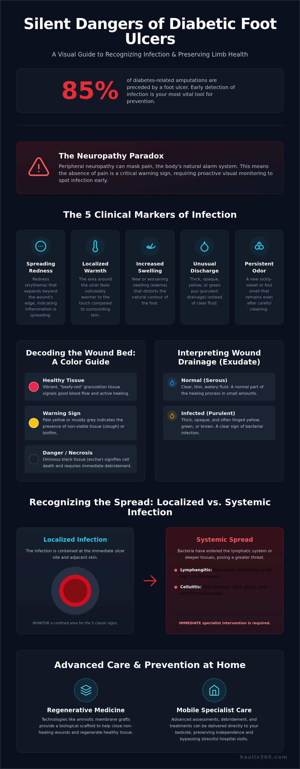
Did you know that 85% of diabetes-related amputations are preceded by a foot ulcer, yet the most critical warning signs often remain completely silent? It's natural to feel a sense of unease when you're managing a non-healing wound at home, especially when you can't rely on pain as a primary guide. When peripheral neuropathy masks the body's typical alarm system, identifying the early signs of infection in diabetic foot ulcer progression becomes your most vital tool for limb preservation.
You deserve the clarity that comes from clinical precision rather than guesswork. This guide will empower you to recognize the subtle markers of bacterial colonization and tissue change, allowing you to bypass the stress of hospital transport and access advanced regenerative care directly at your bedside. We will explore the specific visual indicators defined in the 2026 ADA Standards of Care and explain how data-driven diagnostic tools now allow specialists to treat complex wounds within the comfort of your own home.
Key Takeaways
- Master the "Classic Five" clinical markers of infection, ranging from subtle warmth to specific changes in wound drainage and odor.
- Recognize why the absence of pain is a critical warning sign, requiring a proactive strategy to spot the early signs of infection in diabetic foot ulcer locations.
- Differentiate between localized tissue changes and systemic symptoms to accurately assess the urgency of medical intervention.
- Explore how regenerative technologies, such as amniotic membrane grafts, provide the biological scaffold necessary to close non-healing wounds.
- Learn how mobile specialist care preserves your independence by delivering advanced clinical assessments and debridement directly to your home.
What Are the Early Signs of Infection in Diabetic Foot Ulcer Sites?
A diabetic foot infection represents a critical biological breach where opportunistic bacteria invade the skin and the delicate underlying tissues. In a healthy system, the body’s inflammatory response is a well-orchestrated defense; however, diabetes can mute these signals. Identifying the early signs of infection in diabetic foot ulcer sites requires a disciplined, visual-first approach to compensate for the sensory loss often caused by neuropathy. You aren't just looking for a wound; you're monitoring a complex biological landscape for any shift in its equilibrium.
Clinical mastery of wound care begins with the "Classic Five" markers. These indicators serve as the primary roadmap for your daily assessments. You should remain vigilant for spreading redness, localized warmth that feels distinct from the surrounding skin, and swelling that distorts the foot's natural contours. When these are paired with unusual discharge or a persistent odor, the diagnostic picture becomes clear. Recognizing these early signs of infection in diabetic foot ulcer progression is the first step in moving from passive observation to proactive limb preservation.
The color of the wound bed offers deep insights into the cellular health of the ulcer. A thriving wound displays vibrant, beefy-red granulation tissue, which signals robust oxygen delivery and active healing. If you notice the wound bed transitioning to a pale yellow, muddy grey, or an ominous black, it's likely that bacterial biofilms or tissue death have taken hold. In rare, high-urgency cases, you might even detect crepitus. This is a subtle, crackling sensation under the skin caused by gas bubbles produced by specific bacteria, and it demands immediate specialist evaluation to prevent deep-tissue damage.
Visual Changes and Discoloration
Precision in your visual assessment can prevent a localized issue from becoming a systemic crisis. You must watch for periwound erythema, which is a flush of redness that migrates significantly beyond the ulcer's immediate edge. While healthy granulation tissue looks like textured red velvet, infected slough appears as a tenacious, stringy yellow or white film that effectively stalls the healing process. A sharp demarcation line marks the precise boundary where viable, healthy tissue meets compromised, necrotic cells.
Odor and Exudate Management
Your senses are powerful diagnostic tools when interpreted with clinical rigor. Bacterial colonization often produces a sickly-sweet or putrid scent that lingers even after the wound has been carefully cleansed. It's also essential to differentiate between normal serous fluid, which is clear and thin, and purulent drainage, which is thick, opaque, and often tinged with green or yellow. A sudden, unexplained increase in wound moisture is rarely a neutral event; it's often the body's attempt to flush out deep-seated pathogens that are overwhelming the local environment.
Localized vs. Systemic Infection: Recognizing the Spread
Infection is not a static state; it's a dynamic progression that can move from a localized breach to a systemic threat. A localized infection remains confined to the immediate ulcer site and the adjacent skin. While these are the Clinical Signs of Infection in Diabetic Foot Ulcers that we initially monitor, they can quickly escalate if the body's defenses are compromised. If you observe red streaks extending from the wound upward toward the ankle or leg, this is lymphangitis. It signals that bacteria have entered the lymphatic system and requires immediate specialist intervention to prevent further spread through the body's internal networks.
Underlying vascular health plays a decisive role in how your body manages these biological threats. For many patients, managing edema in legs is a critical component of wound success because fluid retention creates a physical barrier that slows the immune response. Swollen tissue compresses small blood vessels, restricting the delivery of oxygen and white blood cells needed to combat the early signs of infection in diabetic foot ulcer sites. By addressing this fluid buildup, you're not just improving comfort; you're optimizing the biological environment for healing.
Signs of Cellulitis and Deep Tissue Involvement
When an infection penetrates deeper than the surface layers, the skin often takes on a tight, glossy appearance and feels unusually hot to the touch. This isn't just surface-level irritation; it's a sign of cellulitis, an infection of the deeper skin tissues. If left unmanaged, the pathogens can bypass the dermal layers entirely and reach the bone, leading to a complex condition known as osteomyelitis. You should also check for fluctuance, which is a spongy, wave-like feel under the skin that indicates a localized abscess requiring clinical drainage.
Systemic Triggers: When to Seek Urgent Help
A systemic infection occurs when the body's entire inflammatory system is activated to fight a spreading pathogen. You must watch for the "Sepsis Alert" trio: fever, chills, and sudden mental confusion. These symptoms suggest that bacteria may have reached the bloodstream, necessitating urgent medical attention. Additionally, if you notice unexplained spikes in blood sugar levels, it's often a biological signal that the body is under stress from an underlying infection. Precise identification is key to a successful recovery, which is why wound culture and sensitivity testing is essential for tailoring an effective antibiotic strategy. Ensuring these markers are caught early allows for more effective, limb-preserving outcomes, and a bedside wound evaluation can provide the clinical clarity you need without the stress of travel.
The Neuropathy Paradox: Why "No Pain" Is a Warning Sign
Pain is the body's natural alarm system, yet for those managing diabetes, this alarm is often permanently silenced. The most dangerous misconception in wound care is the belief that a lack of pain indicates a lack of infection. Peripheral neuropathy systematically destroys the sensory feedback loop in the lower extremities, meaning the early signs of infection in diabetic foot ulcer sites won't present as the throbbing or soreness you might expect. When nerves are compromised, a deep-seated infection can flourish entirely undetected by your physical senses.
A successful diabetic ulcer treatment plan must account for this sensory deficit by prioritizing objective clinical data over subjective feeling. Since you can't rely on what your foot feels, you must rely on what your eyes see. We recommend the "Mirror Test" for daily inspections. Use a long-handled mirror to examine the plantar surface of the foot, looking for the visual markers of infection discussed earlier. This shift from feeling to seeing is a fundamental pillar of proactive limb preservation and total-body health.
Silent Progression of Diabetic Ulcers
Neuropathy affects up to 50% of adults with diabetes during their lifetime. This widespread condition allows for the silent progression of ulcers, as thick calluses often form over pressure points and act as a biological mask for deep abscesses. It's common for patients to discover a problem only when they see blood or unusual drainage on their socks, by which point the early signs of infection in diabetic foot ulcer tissue may have already transitioned into a more serious stage. If you see any unexpected moisture on your footwear, it's time to look deeper.
When to Call a Glendale or Los Angeles Specialist
Waiting for pain to arrive is a high-risk strategy that often leads to avoidable hospitalizations. If you live in Los Angeles or Glendale, you have access to board-certified specialists who can conduct a thorough bedside evaluation in the comfort of your home. Medicare Part B covers these professional wound assessments, ensuring that financial barriers don't stand in the way of elite care. A mobile assessment provides the diagnostic precision needed to identify "silent" infections before they threaten your mobility. You don't have to wait for a signal that your body can no longer send; take the initiative to secure your long-term vitality through professional intervention.
Advanced Prevention: Regenerative Medicine and Skin Substitutes
Standard wound care often stalls because it fails to address the microscopic environment of a chronic ulcer. Sharp debridement is the clinical necessity of removing infected biofilm, a resilient bacterial shield that prevents antibiotics and the body's own healing cells from reaching the wound bed. By meticulously clearing this debris, a specialist effectively restarts the healing clock, transforming a stagnant wound into an active recovery site. This process is far more sophisticated than simple cleaning; it's a precision biological reset that prepares the tissue for advanced intervention.
Once the wound bed is prepared, advanced regenerative therapies provide the architecture for permanent closure. Utilizing a bio-engineered skin substitute allows clinicians to seal the biological breach before the early signs of infection in diabetic foot ulcer tissue can escalate into a deep-tissue crisis. Additionally, Negative Pressure Wound Therapy (NPWT), commonly known as a Wound VAC, uses controlled suction to stimulate local blood flow and continuously remove excess exudate. This technology is particularly effective for managing high-volume drainage, ensuring the wound environment remains optimized for cellular migration rather than bacterial growth.
The Science of Amniotic Allografts
Amniotic tissue represents the pinnacle of modern regenerative medicine, offering a sophisticated alternative to traditional dressings. These allografts naturally contain a dense concentration of growth factors and cytokines that actively resist bacterial colonization while dampening chronic inflammation. By providing a biological scaffold, these grafts accelerate tissue closure and significantly lower the risk of limb-threatening complications. In Southern California, bedside grafting has become the preferred standard for individuals who prioritize both clinical excellence and the convenience of staying in their own environment.
Infection Mitigation via Debridement
Effective infection mitigation requires more than just a sterile bandage. Clinical debridement performed by a board-certified specialist removes non-viable tissue that serves as a breeding ground for bacteria. We pair this with specialized antimicrobial dressings to maintain a hostile environment for pathogens. By partnering with home health agencies, we ensure every patient receives consistent, expert monitoring that catches the early signs of infection in diabetic foot ulcer sites before they threaten systemic health. If you're ready to move beyond traditional bandages, request a mobile wound care consultation to see if regenerative grafting is right for you.
Mobile Wound Care: Expert Intervention at Your Bedside
The transition from observation to intervention is the most critical phase of limb preservation. Healix360 redefines the recovery experience by bringing board-certified wound specialists directly to your home in Los Angeles or Glendale. This mobile model eliminates the physical trauma often associated with transporting elderly or immobile patients, while simultaneously removing the risk of exposure to hospital-acquired infections. When you identify the early signs of infection in diabetic foot ulcer sites, the most effective response is immediate, professional intervention in a controlled, comfortable environment.
Our specialists often collaborate with families and home health agencies to manage multiple complex issues simultaneously, such as a bed sores ulcer alongside a non-healing diabetic wound. A bedside visit is a comprehensive clinical encounter rather than a simple checkup. It includes sharp debridement to prepare the wound bed, the application of amniotic membrane grafts when indicated, and the development of a personalized care plan tailored to your specific biological needs. This high-touch approach ensures that no detail of your recovery is overlooked, bridging the gap between clinical data and your daily quality of life.
Why Mobile Care is Superior for Diabetic Patients
Mobile care is built on the philosophy of proactive prevention. Instead of a "wait-and-see" approach that often leads to the emergency room, we use immediate, on-site diagnostics to confirm if the early signs of infection in diabetic foot ulcer tissue require a change in antibiotic strategy or advanced grafting. We also conduct personalized assessments of your home environment, identifying potential pressure risks or gait issues that could lead to future ulcers. Because we are a Medicare Part B provider, the billing process remains seamless, allowing you to focus entirely on your physical excellence and long-term vitality.
Next Steps for Southern California Residents
Securing elite wound care in Southern California is a straightforward process designed for high-achievers who value their health as their most valuable asset. You can schedule an urgent bedside evaluation to address concerns before they escalate into systemic complications. Before your first consultation, ensure you have a list of current medications and any recent blood sugar logs ready for review. This data helps our strategists create a clear, actionable roadmap for your recovery. Taking this step today ensures you remain in expert hands, preserving your independence and your future mobility.
Ready to protect your health? Schedule a Mobile Wound Assessment in Southern California
Securing Your Path to Proactive Limb Preservation
Mastery over your health begins with the transition from reactive observation to strategic intervention. By understanding that pain is an unreliable narrator in the presence of neuropathy, you can prioritize the objective visual markers that define the early signs of infection in diabetic foot ulcer progression. Identifying these subtle shifts allows you to bypass the traditional hospital cycle and access the latest breakthroughs in regenerative medicine, such as amniotic membrane grafts, from the comfort of your own home.
As a dedicated Medicare Part B provider, Healix360 serves as your elite guide through the complexities of chronic wound management. Our board-certified specialists bring precision debridement and advanced diagnostic tools directly to your bedside in Los Angeles, Glendale, and across Southern California. You don't have to navigate this journey alone or risk the trauma of unnecessary hospital transport when expert care is available at your door.
Take the first step toward long-term vitality today. Request an Expert Bedside Wound Assessment to ensure your recovery remains on a trajectory of excellence and physical freedom. You deserve a partner who values your mobility as much as you do.
Frequently Asked Questions
Can a diabetic foot ulcer be infected without redness?
Yes, an infection can exist without visible redness, especially if peripheral artery disease or poor circulation limits the body's inflammatory response. In these cases, you must look for other early signs of infection in diabetic foot ulcer sites, such as a sudden increase in drainage or a change in tissue texture. Relying on redness alone can be misleading for those with compromised vascular health.
How fast does an infection spread in a diabetic foot ulcer?
Infection in a diabetic foot can progress with alarming speed, potentially moving from a localized issue to a deep tissue crisis within 24 to 48 hours. Because high blood sugar provides a fuel source for bacteria, the escalation can be much faster than in non diabetic individuals. Immediate specialist evaluation is the most effective way to halt this rapid biological spread and protect your long term mobility.
Does a foul smell always mean my ulcer is infected?
A persistent foul odor is a primary clinical indicator of bacterial colonization and usually signals an active infection. While some specialized dressings can produce a mild scent, a putrid or sickly sweet smell typically means bacteria are actively breaking down tissue. If cleansing the wound doesn't resolve the odor, it's time to seek a professional bedside assessment to prevent further complications.
What color is an infected diabetic foot ulcer?
An infected ulcer often transitions from a healthy pink to shades of yellow, grey, or black. Yellow or white stringy material, known as slough, represents a buildup of dead cells and bacteria that stalls the healing process. Monitoring these color shifts is essential for identifying the early signs of infection in diabetic foot ulcer progression before the damage reaches the underlying bone.
Can I treat a minor ulcer infection at home with over-the-counter creams?
No, you should avoid treating a suspected infection with over the counter creams, as these can trap bacteria and create a moist environment where pathogens thrive. Professional wound care requires clinical debridement to remove biofilms and culture specific antibiotics to target the exact bacteria present. Home based specialist care provides these advanced interventions safely without the risks of self treatment.
Does Medicare Part B cover mobile wound care for diabetic ulcers?
Yes, Medicare Part B typically covers mobile wound care services when they're deemed medically necessary for chronic or non healing ulcers. This coverage includes bedside evaluations, sharp debridement, and advanced regenerative treatments like amniotic grafts. Our team coordinates directly with Medicare to ensure your focus remains on healing rather than billing logistics.
What is the risk of amputation if an ulcer infection is caught early?
The risk of amputation decreases dramatically when an infection is identified and treated in its earliest stages. Clinical data suggests that early intervention is the single most important factor in limb salvage. By catching subtle changes before they reach the bone or bloodstream, we can implement regenerative strategies that preserve your mobility and long term vitality.


